In sum, if it seems that I have simply misunderstood what Kant and deontology are all about,
it's because I am advancing an alternative hypothesis to the standard Kantian/deontological understanding
of what Kant and deontology are all about. I am putting forth an empirical hypothesis
about the hidden psychological essence of deontology, and it cannot be dismissed a priori
for the same reason that tropical islanders cannot know a priori whether ice is a form of water.
- Joshua Greene's (2008, p. 74) 'The secret joke of Kant's soul'
Parts of the Human Brain
The human brain (Holocene epoch)
The brain is divided into 5 parts:
The cerebrum or telencephalon (forebrain);
The cerebellum or metencephalon (hindbrain);
The brain stem;
The pituitary gland;
The hypothalamus
The cerebrum is the largest part of the brain
The cerebral cortex is the outer layer of neural tissue of the cerebrum
The cerebral cortex has an important role in attention, perception, awareness, thought, memory, language, and consciousness
Parts of the Human Brain
GIF Digram
Neural Organization & Function
Cerebrum
The cerebrum is the largest part of the brain
It contains the cerebral cortex
Cerebellum
The cerebellum is a major structure of the hindbrain that is located near the brainstem at the back of the brain
It plays an important role in motor control
Brain stem
The brain stem is the stalk-like part of the brain that connects the cerebrum to the spinal cord
Pituitary gland
The pituitary gland (or 'master gland') is a pea-sized gland at the base of the brain, responsible for producing a number of hormones
Hypothalamus
The hypothalamus is a part of the brain that has a vital role in controlling many bodily functions, including the release of hormones from the pituitary gland
The hypothalamus is part of the limbic system and it is located below the thalamus
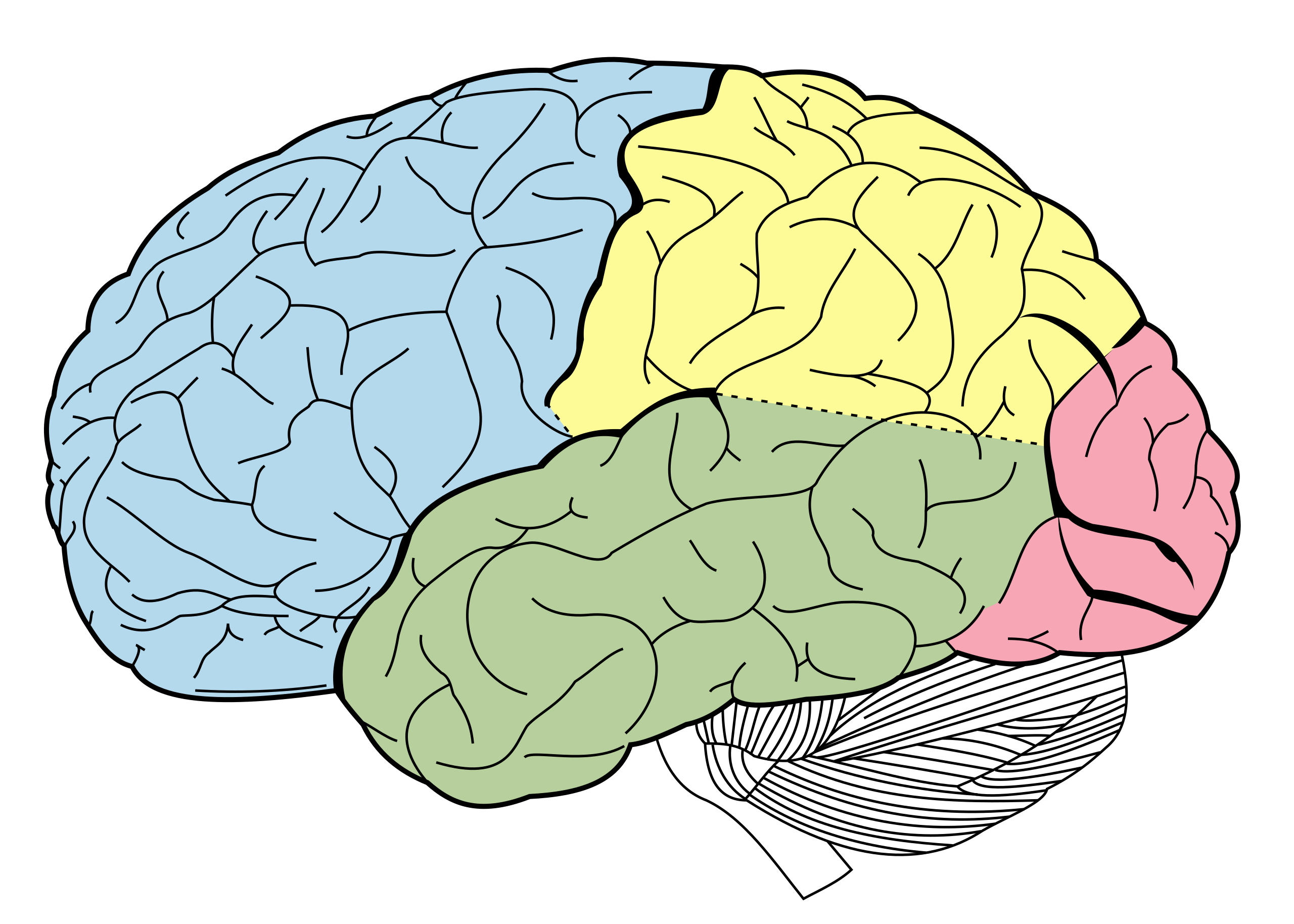
The cerebral cortex
We can divide the cerebral cortex into 4 lobes:
The frontal lobe;
The parietal lobe;
The temporal lobe;
The occipital lobe
Parts of the Cerebral Cortex
GIF Digram
Neural Organization & Function
Frontal lobe
The frontal lobe is located in the forward part of the brain
It is involved in reasoning, motor control, emotion, and language
It contains the following:
The motor cortex (for planning, control, and execution of voluntary movements);
The prefrontal cortex (for higher-level cognitive functions);
Broca's area (for speech production and language processing)
Parietal lobe
The parietal lobe is located above the temporal lobe and behind the frontal lobe
It integrates sensory information among various modalities
It contains the somatosensory cortex, which is essential for processing sensory information from across the body
Temporal lobe
The temporal lobe is associated with hearing, memory, emotion, and certain aspects of language processing
'Temporal' means 'near the head's temples'
The temporal lobe contains the following:
The auditory cortex (for processing auditory information);
The amygdala (Latin for 'almond'), an almond-shaped collection of nuclei within the temporal lobe for processing fearful and threatening stimuli
Wernicke's area (for speech comprehension)
NOTE: While patients with damage to Broca's area have difficulty with language production, patients with damage to Wernicke's area have problems with speech comprehension
Occipital lobe
The occipital lobe is located at the back of the head
'Occipital' derives from the Latin 'ob' (for 'behind') and 'caput' (for 'the head')
It contains the primary visual cortex, which is responsible for interpreting the incoming visual information
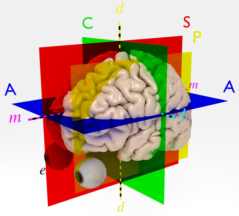
3 anatomical planes for the human brain
Anatomical Planes & Axes of the Human Brain
Diagram
Description
Anatomical axes of the human brain
A denotes the axial, transverse, or horizontal plane
S denotes the sagittal plane, passing between the 2 cerebral hemispheres
C denotes the coronal plane
d denotes the dorsoventral axis, running from the dorsal (superior or toward the scalp) to the ventral (inferior or toward the bottom)
l denotes the lateral axis (away from the middle)
m denotes the medial axis (toward the middle)
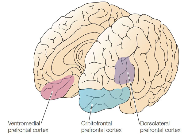
Prefrontal cortex
The ventromedial prefrontal cortex (or VMPFC) is the part of the prefrontal cortex that is located in the frontal lobe, at the ventral (inferior) part of the dorsoventral axis, and along the medial axis (toward the middle)
The dorsolateral prefrontal cortex (or DLPFC) is the part of the prefrontal cortex that is located in the frontal lobe, at the dorsal (superior) part of the dorsoventral axis, and along the lateral axis (away from the middle)
